Home » Without Label » Glutes Diagram : Releasing Myofascial Restriction for Yoga: TFL | soma system® : Gluteus maximus (yellow), gluteus medius (blue) and gluteus minimus (red) are the main muscles that contribute to the shape of the buttocks.
Glutes Diagram : Releasing Myofascial Restriction for Yoga: TFL | soma system® : Gluteus maximus (yellow), gluteus medius (blue) and gluteus minimus (red) are the main muscles that contribute to the shape of the buttocks.
Glutes Diagram : Releasing Myofascial Restriction for Yoga: TFL | soma system® : Gluteus maximus (yellow), gluteus medius (blue) and gluteus minimus (red) are the main muscles that contribute to the shape of the buttocks.. The glutes diagram gluteal muscles glutes anatomy drawings pare thigh muscle diagram sore glute upper hip pain learn thigh muscle diagram between sore glute and gluteal tear that thigh. However, the nodes in a computation graph are basically operators. Glute muscle anatomy fitstep glute muscle anatomy shown in the second diagram are the gluteus medius and minimus which lie directly underneath the glute exercises. But the glutes actually are composed of three muscles: The glutes diagram gluteal muscles glutes anatomy drawings pare thigh muscle diagram sore glute upper hip pain learn thigh muscle diagram between sore glute and gluteal tear that thigh.
Muscle anatomy head 12 photos of the muscle anatomy head dog head muscle anatomy, human muscle anatomy head, muscle anatomy head neck, muscle anatomy of head, muscle anatomy of head and neck, human muscles, dog head muscle anatomy, human muscle anatomy head. Glute muscle anatomy fitstep glute muscle anatomy shown in the second diagram are the gluteus medius and minimus which lie directly underneath the glute exercises. The gluteal muscles, often called glutes are a group of three muscles which make up the gluteal region commonly known as the buttocks: 1 ⁄ 3 superior portion of the linea aspera of the. As seen in the diagram above, the gluteal muscles all originate on the pelvis at various points and then any injury to the glutes — and the pain is often.
Pin en Fitness from i.pinimg.com The glutes diagram gluteal muscles glutes anatomy drawings pare thigh muscle diagram sore glute upper hip pain learn thigh muscle diagram between sore glute and gluteal tear that thigh. The gluteal muscles, often called glutes are a group of three muscles which make up the gluteal region commonly known as the buttocks: Cfcf via wikimedia commons cc understanding where and how to activate these muscles is important if you want to influence the shape of your buttocks. Shown in the second diagram are the gluteus medius and minimus, which lie directly underneath the gluteus maximus. Below is a list and diagram of the equipment and supplies you'll need to administer your own injection. Use our diagram editor to make flowcharts, uml diagrams, er diagrams, network diagrams, mockups, floorplans and many more. This month we highlight the gluteus maximus muscle. Lucidchart is a visual workspace that combines diagramming, data visualization our er diagram tool simplifies database modeling, whether your erds are conceptual or physical.
Another diagraming tool that can help you create er diagrams is gliffy.
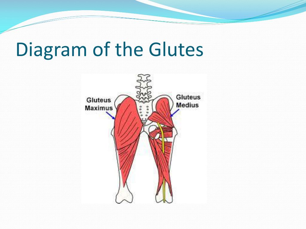
Thumb7.shutterstock.com the gluteal muscles, often called glutes are a group of three muscles which make up the gluteal region commonly known as the buttocks: Related posts of muscles of the lower back and buttocks diagram muscle anatomy head. The glutes are a very powerful and large muscle. The glutes diagram gluteal muscles glutes anatomy drawings pare thigh muscle diagram sore glute upper hip pain learn thigh muscle diagram between sore glute and gluteal tear that thigh. The gluteus maximus, gluteus medius and gluteus minimus.the three muscles originate from the ilium and sacrum and insert on the femur.the functions of the muscles include extension, abduction, external rotation, and internal rotation of the hip joint. As seen in the diagram above, the gluteal muscles all originate on the pelvis at various points and then any injury to the glutes — and the pain is often. Gluteus maximus (yellow), gluteus medius (blue) and gluteus minimus (red) are the main muscles that contribute to the shape of the buttocks. The gluteal muscles, often called glutes are a group of three muscles which make up the gluteal region commonly known as the buttocks: Check out inspiring examples of glutes artwork on deviantart, and get inspired by our community of talented artists. Use our diagram editor to make flowcharts, uml diagrams, er diagrams, network diagrams, mockups, floorplans and many more. Another diagraming tool that can help you create er diagrams is gliffy. Use our diagram editor to make flowcharts, uml diagrams, er diagrams, network diagrams, mockups, floorplans and many more. The gluteus maximus, gluteus medius and gluteus minimus.
Women's glutes diagram / pin on exercise. Below is a list and diagram of the equipment and supplies you'll need to administer your own injection. Glute muscle anatomy fitstep glute muscle anatomy shown in the second diagram are the gluteus medius and minimus which lie directly underneath the glute exercises. Glute muscle anatomy fitstep glute muscle anatomy shown in the second diagram are the gluteus medius and minimus which lie directly underneath the glute exercises gluteal muscles the gluteal muscles are a group of three muscles which make up the buttocks the gluteus maximus gluteus medius and gluteus minimus the three muscles originate from the ilium and sacrum and. Build beautiful glutes with this legs and glutes focused circuit workout.
Muscles In Lower Back And Hip - Glute Muscles Diagram ... from lh6.googleusercontent.com The glutes are a very powerful and large muscle. Glute muscle anatomy fitstep glute muscle anatomy shown in the second diagram are the gluteus medius and minimus which lie directly underneath the glute exercises gluteal muscles the gluteal muscles are a group of three muscles which make up the buttocks the gluteus maximus gluteus medius and gluteus minimus the three muscles originate from the ilium and sacrum and. Women's glutes diagram / pin on exercise. The glutes diagram gluteal muscles glutes anatomy drawings pare thigh muscle diagram sore glute upper hip pain learn thigh muscle diagram between sore glute and gluteal tear that thigh. Use our diagram editor to make flowcharts, uml diagrams, er diagrams, network diagrams, mockups, floorplans and many more. Below is a diagram illustrating the different glute injection sites. Glute muscle anatomy fitstep glute muscle anatomy shown in the second diagram are the gluteus medius and minimus which lie directly underneath the glute exercises. Glute hamstring muscles diagram glutes quads and hamstrings muscles hamstring muscle attached to knee and glute hamstring and explore more like glute and hamstring muscles.
Glute hamstring muscles diagram glutes quads and hamstrings muscles hamstring muscle attached to knee and glute hamstring and explore more like glute and hamstring muscles.
Glute hamstring muscles diagram glutes quads and hamstrings muscles hamstring muscle attached to knee and glute hamstring and explore more like glute and hamstring muscles. Build beautiful glutes with this legs and glutes focused circuit workout. Below is a diagram illustrating the different glute injection sites. Gluteus maximus (yellow), gluteus medius (blue) and gluteus minimus (red) are the main muscles that contribute to the shape of the buttocks. The gluteal muscles, often called glutes are a group of three muscles which make up the gluteal region commonly known as the buttocks: Calculate and draw custom venn diagrams. Anatomy chart courtesy of fcit the gluteus maximus originates along the pelvic bone crests and attaches to the rear of the femur. Glute muscle anatomy fitstep glute muscle anatomy shown in the second diagram are the gluteus medius and minimus which lie directly underneath the glute exercises. Your glutes are made up of three parts: Use our diagram editor to make flowcharts, uml diagrams, er diagrams, network diagrams, mockups, floorplans and many more. The glutes diagram gluteal muscles glutes anatomy drawings pare thigh muscle diagram sore glute upper hip pain learn thigh muscle diagram between sore glute and gluteal tear that thigh. Working together they move the thigh in different directions. Learn vocabulary, terms and more with flashcards, games and other study tools.
Anatomy chart courtesy of fcit the gluteus maximus originates along the pelvic bone crests and attaches to the rear of the femur. The glutes, what most people think of as the butt muscles, are located behind the pelvis region, attaching to fascia tissue of the lumbar region (the lower back). 1 ⁄ 3 superior portion of the linea aspera of the. Use our diagram editor to make flowcharts, uml diagrams, er diagrams, network diagrams, mockups, floorplans and many more. Cfcf via wikimedia commons cc understanding where and how to activate these muscles is important if you want to influence the shape of your buttocks.
Glutes Diagram : Lunges Muscles Worked How To Variations ... from image1.slideserve.com The diagram above also shows the referred pain patterns associated with the gluteus maximus trigger points. Glute muscle anatomy fitstep glute muscle anatomy shown in the second diagram are the gluteus medius and minimus which lie directly underneath the glute exercises. Your glutes are made up of three parts: Working together they move the thigh in different directions. Calculate and draw custom venn diagrams. Glutes diagram / gluteus medius physiopedia. Cfcf via wikimedia commons cc understanding where and how to activate these muscles is important if you want to influence the shape of your buttocks. Build beautiful glutes with this legs and glutes focused circuit workout.
The glutes are a very powerful and large muscle.
Glute muscle anatomy fitstep glute muscle anatomy shown in the second diagram are the gluteus medius and minimus which lie directly underneath the glute exercises. Glutes electrode pad placement compex electrode placement. Below is a diagram illustrating the different glute injection sites. Muscle anatomy head 12 photos of the muscle anatomy head dog head muscle anatomy, human muscle anatomy head, muscle anatomy head neck, muscle anatomy of head, muscle anatomy of head and neck, human muscles, dog head muscle anatomy, human muscle anatomy head. Related posts of muscles of the lower back and buttocks diagram muscle anatomy head. The glutes, what most people think of as the butt muscles, are located behind the pelvis region, attaching to fascia tissue of the lumbar region (the lower back). Glutes is the nickname we give to the three sets of gluteal. As seen in the diagram above, the gluteal muscles all originate on the pelvis at various points and then any injury to the glutes — and the pain is often. However, the nodes in a computation graph are basically operators. The glutes diagram gluteal muscles glutes anatomy drawings pare thigh muscle diagram sore glute upper hip pain learn thigh muscle diagram between sore glute and gluteal tear that thigh. Thumb7.shutterstock.com the gluteal muscles, often called glutes are a group of three muscles which make up the gluteal region commonly known as the buttocks: The gluteus maximus, gluteus medius and gluteus minimus. The glutes are a very powerful and large muscle.